PDF Publication Title:
Text from PDF Page: 011
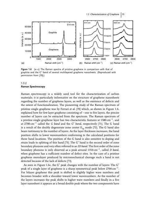
(a) (b) 2800 2600 2700 2800 (c) Raman shift (cm−1) 1500 2000 2500 3000 Raman shift (cm−1) 2600 2700 Raman shift (cm−1) Graphite Graphene Graphite 10 layers 5 layers 2 layers 1 layer 514 nm 50 000 40 000 30 000 20 000 10 000 0 1.3.2 Raman Spectroscopy Raman spectroscopy is a widely used tool for the characterization of carbon materials; it is particularly informative on the structure of graphene nanosheets regarding the number of graphene layers, as well as the existence of defects and the extent of functionalization. The pioneering study of the Raman spectrum of pristine single graphene was by Ferrari et al. [70] which, as shown in Figure 1.6, explained how for few-layer graphene consisting of – one to five layers, the precise number of layers can be extracted from the spectrum. The Raman spectrum of a pristine single graphene layer has two characteristic features at 1580cm−1, and at 2700cm−1 called the G band and the G′ band, respectively [71]. The G band is a result of the doubly degenerate zone center E2g mode [72]. The G band also bears testimony to the number of layers. As the layer thickness increases, the band position shifts to lower wavenumbers conforming to the calculated positions for these band locations. The position of the G band is also sensitive to doping and strain leads to splitting of this band [73]. The G′ band is the second order of zone boundary phonons and very often referred to as 2D band. The first order of the zone boundary phonons is only observed as a peak around 1350cm−1, called D band, when graphene has a sufficient number of defect sites. In the case of a pristine graphene monolayer produced by micromechanical cleavage such a band is not detected because of the lack of defects [73]. As seen in Figure 1.6c, the G′ peak changes with the number of layers: The G′ peak of a single layer of graphene is a sharp symmetrical peak below 2700cm−1. For bilayer graphene this peak is shifted to slightly higher wave numbers and becomes broader with a shoulder toward lower wavenumbers. As the number of the layers increases the peak shifts to higher wave numbers and finally in a five layer nanosheet it appears as a broad double peak where the two components have (a–c) The Raman spectra of pristine graphene in comparison with that of graphite and the G′ band of several multilayered graphene nanosheets. (Reproduced with permission from [70].) Figure 1.6 1.3 Characterization of Graphene 11 514 nm 633 nm Intensity (a. u.) Intensity (a. u.)PDF Image | An Introduction to Graphene
PDF Search Title:
An Introduction to GrapheneOriginal File Name Searched:
352733551X-c01.pdfDIY PDF Search: Google It | Yahoo | Bing
Salgenx Redox Flow Battery Technology: Power up your energy storage game with Salgenx Salt Water Battery. With its advanced technology, the flow battery provides reliable, scalable, and sustainable energy storage for utility-scale projects. Upgrade to a Salgenx flow battery today and take control of your energy future.
| CONTACT TEL: 608-238-6001 Email: greg@infinityturbine.com | RSS | AMP |