PDF Publication Title:
Text from PDF Page: 009
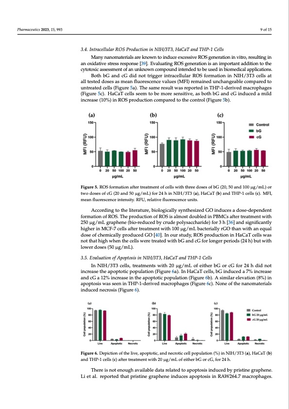
Pharmaceutics 2023, 15, x FOR PEER REVIEW 9 of 16 Pharmaceutics 2023, 15, x FOR PEER REVIEW 9 of 16 Pharmaceutics 2023, 15, 993 of cells (~30% reduction), the magnitude of this effect was lower than that exerted cG of cells (~30% reduction), the magnitude of this effect was lower than that exerted cG doses higher than 50 μg/mL. Although bG at 100 μg/mL reduced the long-term survival doses higher than 50 μg/mL. Although bG at 100 μg/mL reduced the long-term survival (~50%) with a half-dose (50 μg/mL). 9 of 15 (~50%) with a half-dose (50 μg/mL). 3.4. Intracellular ROS Production in NIH/3T3, HaCaT and THP-1 Cells 3.4. Intracellular ROS Production in NIH/3T3, HaCaT and THP-1 Cells 3.4. Intracellular ROS Production in NIH/3T3, HaCaT and THP-1 Cells Many nanomaterials are known to induce excessive ROS generation in vitro, result- Many nanomaterials are known to induce excessive ROS generation in vitro, result- Many nanomaterials are known to induce excessive ROS generation in vitro, resulting in ing in an oxidative stress response [39]. Evaluating ROS generation is an important addi- ing in an oxidative stress response [39]. Evaluating ROS generation is an important addi- an oxidative stress response [39]. Evaluating ROS generation is an important addition to the tion to the cytotoxic assessment of an unknown compound intended to be used in bio- tion to the cytotoxic assessment of an unknown compound intended to be used in bio- cytotoxic assessment of an unknown compound intended to be used in biomedical applications. medical applications. medical applications. Both bG and cG did not trigger intracellular ROS formation in NIH/3T3 cells at Both bG and cG did not trigger intracellular ROS formation in NIH/3T3 cells at all Both bG and cG did not trigger intracellular ROS formation in NIH/3T3 cells at all all tested doses as mean fluorescence values (MFI) remained unchangeable compared to tested doses as mean fluorescence values (MFI) remained unchangeable compared to un- tested doses as mean fluorescence values (MFI) remained unchangeable compared to un- untreated cells (Figure 5a). The same result was reported in THP-1-derived macrophages treated cells (Figure 5a). The same result was reported in THP-1-derived macrophages treated cells (Figure 5a). The same result was reported in THP-1-derived macrophages (Figure 5c). HaCaT cells seem to be more sensitive, as both bG and cG induced a mild (Figure 5c). HaCaT cells seem to be more sensitive, as both bG and cG induced a mild (Figure 5c). HaCaT cells seem to be more sensitive, as both bG and cG induced a mild increase (10%) in ROS production compared to the control (Figure 5b). increase (10%) in ROS production compared to the control (Figure 5b). increase (10%) in ROS production compared to the control (Figure 5b). Figure 5. ROS formationaftertreatmenttofcelllswiitththredosesofffbG(2(20,,,550aand11000μμμgg/m/mLL))oorr two doses ofcG(20and50μμg//mL))fforr24hiinNIIH/33T33((a(a)),),HaaCaaTT(b(b))aannddTTHPP-1-1cceellsls(c(c)c.).MFFI,I, meanfluorescenceintensiitty..RFU,,rrellattiivefflluorreesscceencceeunnititss.. mean fluorescence intensity. RFU, relative fluorescence units. According to the literature, biologicalllysyntthessiizeedGOiininducceessadossee--d-deepeennddeenntt formationoffROSS..TThhehepprpordodudcutcicottiinononfoRffORSOiSsSaiislsmaalolmstodsstotudboolueubdblelienddPinBinMPPBCBMsMaCfCtsesraafttfreteerartmrtereanatmtmweenitntht 2w5i0thμ2g5/0mμLg/g/mraLpghgreranpeh(ebenioe-(r(bebidiou--rcredubcyedcrbubydcecrrpuodleyepspaoclclyyhssaarccicdchheaa)rrifdiodree)3)fofhor[r336h]ha[3[n36d6]]saaingndndisfisigcigannifitifl-iy- hcaignhtleyrhinigMheCrFin-7MceCllFs-7a7fctelrllstsraeafafttemrtetrnetatwtmitehnt1tw0w0iitμthg1/100m00Lμgbg/a/mctLeLrbibaacllctyeteriGaialOlylytrhrGaGOnOwthtihatahnnawnitiehtqhauananl deqousealodfocsheomofichaellmyipcaraolldyupcreoduGucOed[4G0O]O. I[[4n40o]].u.IrInsotoudrryss,ttuRudOdyyS,,RpROrOoSSdpuprcrotodidounucctiitnoionHnianiCnHaHTaaCcCaeaTllTsccewellasls nwoatsthnaoththihgaht whihgehnwthencnetlthlhsewcerllelstwrweeaertredttrrweaiattheedbGwiaittnhdbbcGaafnondrdlcocGnGgfefoorrploleonrnigogedersrp(p2ee4rirhoio)ddbssu(2t(24w4hith)h) lbouwt ewridthosloeowsw(e5r0dμogs/esms (L50)0. μg//mL)).. 3.5. Evaluation of Apoptosis in NIH/3T3, HaCaT and THP-1 Cells 3.5.EvaluationofApoptosiisiinNIIH//3T3,,HaaCaaTaanddTHP--11Ceellsls In NIH/3T3 cells, treatments with 20 μg/mL of either bG or cG for 24 h did not In NIH/3T3 cells,, treattmenttswiitth20μg//mLoffeeiittheerrbbGoorrccGfoforr2244hhddididnnoottinin-- increase the apoptotic population (Figure 6a). In HaCaT cells, bG induced a 7% increase crease the apoptoticpopullattiion((Fiigurre6a))..IInHaaCaaTcceellsls,,bbGinindduucceeddaa77%ininccrereaasseeaanndd and cG a 12% increase in the apoptotic population (Figure 6b). A similar elevation (8%) in cG a 12% increase in the apoptotic population (Figure 6b). A similar elevation (8%) in cG a 12% increase in the apoptotic population (Figure 6b). A similar elevation (8%) in apoptosis was seen in THP-1-derived macrophages (Figure 6c). None of the nanomaterials apoptosis was seen in THP-1-derived macrophages (Figure 6c). None of the nanomaterials apoptosis was seen in THP-1-derived macrophages (Figure 6c). None of the nanomaterials induced necrosis (Figure 6). induced necrosis (Figure 6). induced necrosis (Figure 6). Figure 6. Depiction of the live, apoptotic, and necrotic cell population (%) in NIH/3T3 (a), HaCaT (b) and THP-1 cells (c) after treatment with 20 μg/mL of either bG or cG, for 24 h. There is not enough available data related to apoptosis induced by pristine graphene. Li et al. reported that pristine graphene induces apoptosis in RAW264.7 macrophages.PDF Image | Green Exfoliation of Graphene
PDF Search Title:
Green Exfoliation of GrapheneOriginal File Name Searched:
pharmaceutics-15-00993.pdfDIY PDF Search: Google It | Yahoo | Bing
Salgenx Redox Flow Battery Technology: Power up your energy storage game with Salgenx Salt Water Battery. With its advanced technology, the flow battery provides reliable, scalable, and sustainable energy storage for utility-scale projects. Upgrade to a Salgenx flow battery today and take control of your energy future.
CONTACT TEL: 608-238-6001 Email: greg@infinityturbine.com (Standard Web Page)