PDF Publication Title:
Text from PDF Page: 005
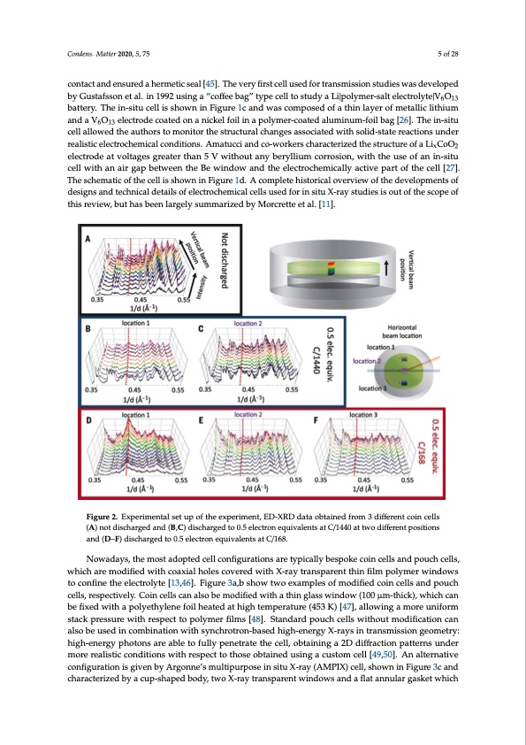
Condens. Matter 2020, 5, 75 5 of 28 contact and ensured a hermetic seal [45]. The very first cell used for transmission studies was developed by Gustafsson et al. in 1992 using a “coffee bag” type cell to study a Li|polymer-salt electrolyte|V6O13 battery. The in-situ cell is shown in Figure 1c and was composed of a thin layer of metallic lithium and a V6O13 electrode coated on a nickel foil in a polymer-coated aluminum-foil bag [26]. The in-situ cell allowed the authors to monitor the structural changes associated with solid-state reactions under realistic electrochemical conditions. Amatucci and co-workers characterized the structure of a LixCoO2 electrode at voltages greater than 5 V without any beryllium corrosion, with the use of an in-situ cell with an air gap between the Be window and the electrochemically active part of the cell [27]. The schematic of the cell is shown in Figure 1d. A complete historical overview of the developments of designs and technical details of electrochemical cells used for in situ X-ray studies is out of the scope of this review, but has been largely summarized by Morcrette et al. [11]. Condens. Matter 2020, 5, x 5 of 30 Figure 2. Experimental set up of the experiment, ED-XRD data obtained from 3 different coin cells Figure 2. Experimental set up of the experiment, ED-XRD data obtained from 3 different coin cells (A) not discharged and (B,C) discharged to 0.5 electron equivalents at C/1440 at two different (A) not discharged and (B,C) discharged to 0.5 electron equivalents at C/1440 at two different positions positions and (D,E,F) discharged to 0.5 electron equivalents at C/168. and (D–F) discharged to 0.5 electron equivalents at C/168. NOonweaodfatyhse,ftihrsetmino-stitaudXoRpDtedceclelsllwcoansfidgeuvrealotipoends ainre1t9y7p8icbayllCyhbieasnpeollkieancodinhicsecllos-auntdhoprosufcohrctehlels, wsthuidchyaorfeamLoid/TifiiSe2dcwelilt.hTchoeaxceiallhwoalsesdceosvigenredwfoirthaXc-ornayvetnratinosnpaalrBenratgtgh–inBrfielnmtanpolydmifferracwtoimndeotewrs twoocorknifinngeinthrefelelecctitornolmytoed[e1,3c,4o6m].pFoisgeudroef3aa5,b0μshMo-wthtiwckoBexwaminpdloews,oafnmdoadTifiefeldonc/oailnumceilnlusmanbdopdoyuinch caellpsa,raelslpelecptilvateelyc.oCnofingucrealltsiocnan[2a5ls].oTbheemsocdheifimeadtiwciothfathtehicneglllaissswhoinwdnowin(1F0ig0uμrme-1tbh.icTkh),ewahuitchocrasn bme ofinxietodrweditthea pdoisloyredtherylienntehfeoTilihS2eactaetdhoadtehdiguhritnegmdpiesrcahtaurrgee,(4w53hiKch) [w47a]s, allsotewpi-ncghanmgeoirne ubantitfeorrym srteascekarpcrhesastutrheawttitmher,esipneccet,tinotphoelypmreevriofiulmswso[4r8k]s.,eSlteacntrdoadredspwoeurcehsecpelalrsawteidthaonudtamnoaldyizfiecdateiox-nsictuan before and after the electrochemical polarization [44]. However, this preliminary cell was not exempt also be used in combination with synchrotron-based high-energy X-rays in transmission geometry: from limitations; only 10% of the electrode was analyzed, due to the penetration depth of the X-rays high-energy photons are able to fully penetrate the cell, obtaining a 2D diffraction patterns under through the cell window (50 μ M-thick) and the electrode (700 μ M-thick). After this first experiment, more realistic conditions with respect to those obtained using a custom cell [49,50]. An alternative a large series of cell configurations were tested by different groups. Dahn and co-workers analyzed configuration is given by Argonne’s multipurpose in situ X-ray (AMPIX) cell, shown in Figure 3c and structural changes in LixTiS2 using an in-situ cell composed of two stainless steel cases, each one with characterized by a cup-shaped body, two X-ray transparent windows and a flat annular gasket which a Be window coated with the electrode. An electrolyte-soaked separator and a polypropylene gasket prevented electrical contact and ensured a hermetic seal [45]. The very first cell used for transmission studies was developed by Gustafsson et al. in 1992 using a “coffee bag” type cell to study a Li|polymer-salt electrolyte|V6O13 battery. The in-situ cell is shown in Figure 1c and was composed of a thin layer of metallic lithium and a V6O13 electrode coated on a nickel foil in a polymer-coated aluminum-foil bag [26]. The in-situ cell allowed the authors to monitor the structural changesPDF Image | Synchrotron-Based X-ray Diffraction for Lithium-Ion Batteries
PDF Search Title:
Synchrotron-Based X-ray Diffraction for Lithium-Ion BatteriesOriginal File Name Searched:
condensedmatter-05-00075.pdfDIY PDF Search: Google It | Yahoo | Bing
Sulfur Deposition on Carbon Nanofibers using Supercritical CO2 Sulfur Deposition on Carbon Nanofibers using Supercritical CO2. Gamma sulfur also known as mother of pearl sulfur and nacreous sulfur... More Info
CO2 Organic Rankine Cycle Experimenter Platform The supercritical CO2 phase change system is both a heat pump and organic rankine cycle which can be used for those purposes and as a supercritical extractor for advanced subcritical and supercritical extraction technology. Uses include producing nanoparticles, precious metal CO2 extraction, lithium battery recycling, and other applications... More Info
| CONTACT TEL: 608-238-6001 Email: greg@infinityturbine.com | RSS | AMP |