PDF Publication Title:
Text from PDF Page: 012
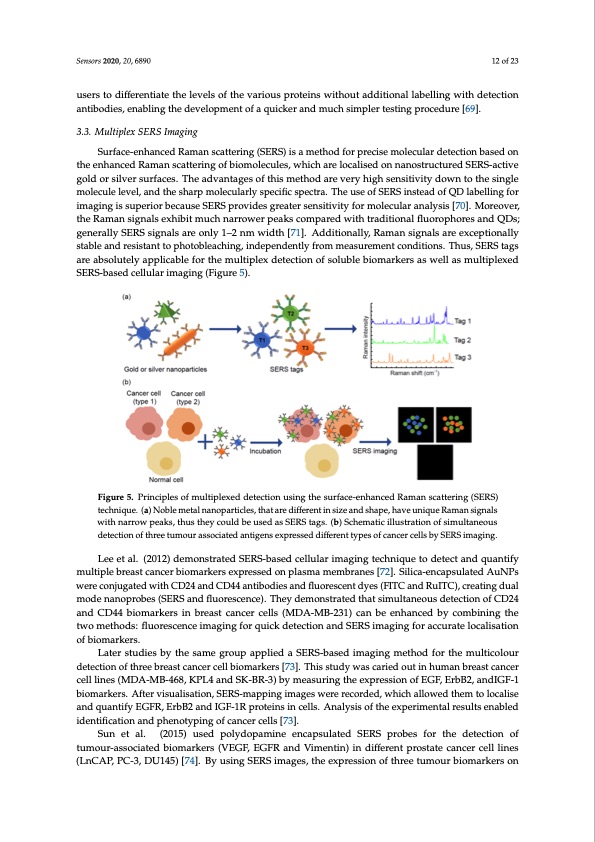
Sensors 2020, 20, 6890 12 of 23 users to differentiate the levels of the various proteins without additional labelling with detection antibodies, enabling the development of a quicker and much simpler testing procedure [69]. 3.3. Multiplex SERS Imaging Surface-enhanced Raman scattering (SERS) is a method for precise molecular detection based on the enhanced Raman scattering of biomolecules, which are localised on nanostructured SERS-active gold or silver surfaces. The advantages of this method are very high sensitivity down to the single molecule level, and the sharp molecularly specific spectra. The use of SERS instead of QD labelling for imaging is superior because SERS provides greater sensitivity for molecular analysis [70]. Moreover, the Raman signals exhibit much narrower peaks compared with traditional fluorophores and QDs; generally SERS signals are only 1–2 nm width [71]. Additionally, Raman signals are exceptionally stable and resistant to photobleaching, independently from measurement conditions. Thus, SERS tags are absolutely applicable for the multiplex detection of soluble biomarkers as well as multiplexed SeEnRsoSrs-2b0a2s0e,d20,cxelFlOuRlaPrEiEmRaRgEiVnIgEW(Figure5). 12of23 Figure 5. Principles of multiplexed detection using the surface-enhanced Raman scattering (SERS) Figure 5. Principles of multiplexed detection using the surface-enhanced Raman scattering (SERS) technique. (a) Noble metal nanoparticles, that are different in size and shape, have unique Raman signals technique. (a) Noble metal nanoparticles, that are different in size and shape, have unique Raman with narrow peaks, thus they could be used as SERS tags. (b) Schematic illustration of simultaneous signals with narrow peaks, thus they could be used as SERS tags. (b) Schematic illustration of detection of three tumour associated antigens expressed different types of cancer cells by SERS imaging. simultaneous detection of three tumour associated antigens expressed different types of cancer cells by SERS imaging. Lee et al. (2012) demonstrated SERS-based cellular imaging technique to detect and quantify multiple breast cancer biomarkers expressed on plasma membranes [72]. Silica-encapsulated AuNPs Lee et al. (2012) demonstrated SERS-based cellular imaging technique to detect and quantify were conjugated with CD24 and CD44 antibodies and fluorescent dyes (FITC and RuITC), creating dual multiple breast cancer biomarkers expressed on plasma membranes [72]. Silica-encapsulated AuNPs mode nanoprobes (SERS and fluorescence). They demonstrated that simultaneous detection of CD24 were conjugated with CD24 and CD44 antibodies and fluorescent dyes (FITC and RuITC), creating and CD44 biomarkers in breast cancer cells (MDA-MB-231) can be enhanced by combining the dual mode nanoprobes (SERS and fluorescence). They demonstrated that simultaneous detection of two methods: fluorescence imaging for quick detection and SERS imaging for accurate localisation CD24 and CD44 biomarkers in breast cancer cells (MDA-MB-231) can be enhanced by combining the of biomarkers. two methods: fluorescence imaging for quick detection and SERS imaging for accurate localisation of Later studies by the same group applied a SERS-based imaging method for the multicolour biomarkers. detection of three breast cancer cell biomarkers [73]. This study was caried out in human breast cancer Later studies by the same group applied a SERS-based imaging method for the multicolour cell lines (MDA-MB-468, KPL4 and SK-BR-3) by measuring the expression of EGF, ErbB2, andIGF-1 detection of three breast cancer cell biomarkers [73]. This study was caried out in human breast biomarkers. After visualisation, SERS-mapping images were recorded, which allowed them to localise cancer cell lines (MDA-MB-468, KPL4 and SK-BR-3) by measuring the expression of EGF, ErbB2, and quantify EGFR, ErbB2 and IGF-1R proteins in cells. Analysis of the experimental results enabled andIGF-1 biomarkers. After visualisation, SERS-mapping images were recorded, which allowed identification and phenotyping of cancer cells [73]. them to localise and quantify EGFR, ErbB2 and IGF-1R proteins in cells. Analysis of the experimental Sun et al. (2015) used polydopamine encapsulated SERS probes for the detection of results enabled identification and phenotyping of cancer cells [73]. tumour-associated biomarkers (VEGF, EGFR and Vimentin) in different prostate cancer cell lines Sun et al. (2015) used polydopamine encapsulated SERS probes for the detection of tumour- (LnCAP, PC-3, DU145) [74]. By using SERS images, the expression of three tumour biomarkers on associated biomarkers (VEGF, EGFR and Vimentin) in different prostate cancer cell lines (LnCAP, PC-3, DU145) [74]. By using SERS images, the expression of three tumour biomarkers on the surface of prostate cancer cells were evaluated visually and three prostate cancer cell lines had been distinguished from each other. The authors suggest that usage of the multiplexed SERS imaging technique could improve the identification of cancer cell phenotypes [74]. Li et al. (2018) developed a multiplexed nanobiosensor by adapting the sandwich-type immunoassay and SERS methods for the detection of cancer biomarkers. Three soluble cancer proteinPDF Image | Multiplexed Nanobiosensors: Current Trends in Early Diagnostics
PDF Search Title:
Multiplexed Nanobiosensors: Current Trends in Early DiagnosticsOriginal File Name Searched:
sensors-20-06890-v2.pdfDIY PDF Search: Google It | Yahoo | Bing
Turbine and System Plans CAD CAM: Special for this month, any plans are $10,000 for complete Cad/Cam blueprints. License is for one build. Try before you buy a production license. More Info
Waste Heat Power Technology: Organic Rankine Cycle uses waste heat to make electricity, shaft horsepower and cooling. More Info
All Turbine and System Products: Infinity Turbine ORD systems, turbine generator sets, build plans and more to use your waste heat from 30C to 100C. More Info
CO2 Phase Change Demonstrator: CO2 goes supercritical at 30 C. This is a experimental platform which you can use to demonstrate phase change with low heat. Includes integration area for small CO2 turbine, static generator, and more. This can also be used for a GTL Gas to Liquids experimental platform. More Info
Introducing the Infinity Turbine Products Infinity Turbine develops and builds systems for making power from waste heat. It also is working on innovative strategies for storing, making, and deploying energy. More Info
Need Strategy? Use our Consulting and analyst services Infinity Turbine LLC is pleased to announce its consulting and analyst services. We have worked in the renewable energy industry as a researcher, developing sales and markets, along with may inventions and innovations. More Info
Made in USA with Global Energy Millennial Web Engine These pages were made with the Global Energy Web PDF Engine using Filemaker (Claris) software.
Infinity Turbine Developing Spinning Disc Reactor SDR or Spinning Disc Reactors reduce processing time for liquid production of Silver Nanoparticles.
| CONTACT TEL: 608-238-6001 Email: greg@infinityturbine.com | RSS | AMP |